Bone Fracture
Even though the bones are strong, they are also susceptible to fractures or breaks. Fractures may be classified based on the
(i) Positioning of the bone ends
(ii) completeness of the break
(iii) orientation of the break relative to the long axis of the bone and
(iv) penetration through the skin. In addition to the above classifications, all fractures can be described in terms of the location of the fracture, the external appearance of the fracture or the nature of the break (Figure 9.12).
The following are the common types of fractures,
-
Tranverse - A fracture that is at right angle to the bone’s long axis.
-
Oblique non-displaced -A fracture that is diagonal to the bone’s long axis and the fractured bone is not displaced from its position.
-
Oblique displaced - A fracture that is diagonal to the bone’s long axis and the fractured bone is displaced from its position.
-
Spiral - Ragged break occurs when excessive twisting forces are applied to a bone (common sports fracture).
-
Greenstick - Bone breaks incompletely, just like a green twig breaks. It is common in children, because of the flexibility of the bones.
-
Comminuted – Bone fragmented into three or more pieces. Particularly common in the aged, whose bones are brittle (hard but easily broken).
Mechanism and healing of a bone fracture
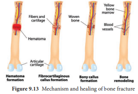
Bone is a cellular, living tissue capable of growth, self-repair and remodeling in response to physical stresses. In the adult skeleton, bone deposit and bone resorption occur. These two processes together constitute in remodeling of bone. There are four major stages in repairing a simple fracture (Figure 9.13).
-
Formation of haematoma When a bone breaks the blood vessels in the bone and surrounding tissues are torn and results in haemorrhage. Due to this a haematoma, a mass of clotted blood forms at the fracture site. The tissues at the site becomes swollen, painful and inflammed. The death of bone cells, occur due to lack of nutrition.
-
Formation of fibrocartilaginous callus Within a few days several events lead to the formation of soft granulation tissue called callus. Capillaries grow into the haematoma and phagocytic cells invade the area and begin to clean up the debris. Meanwhile the fibroblasts and osteoblasts invade from the nearby periosteum and endosteum and begin reconstructing of the bone. The fibroblasts produce fibres. The chondroblasts secrete the cartilage matrix. Within this repair tissue, osteoblasts begin forming spongy bone. The cartilage matrix later calcifies and forms the fibrocartilaginous callus.
-
Formation of Bony callus New bone trabeculae begin to appear in the fibro cartilaginous callus. Gradually that is converted into a bony (hard) callus of spongy bone. Bony callus formation continues until a firm union is formed about two months later to an year for complete woven bone formation.
-
Remodeling of Bone Bony callus formation will be continued for several months. After that the bony callus is remodelled. The excess material on the diaphysis exterior and within the medullary cavity is removed and the compact bone is laid down to reconstruct the shaft walls. The final structure of the remodelled area resembles like the unbroken bony region.