Chapter 6 Microbial Nutrition and Growth Chapter Outline
6.1 Microbial Nutrients 6.2 Nutrient Requirement of
Microorganisms 6.3 Nutritional Types of
Microorganisms 6.4 Photosynthesis 6.5 Microbial Growth 6.6 Measurement of Microbial Growth
org sim
After studying this chapter the student will be able, • To know the essential nutrients
required by bacterial cell. • To differentiate between macronu-
trients and micronutrients. • To describe an organism based on
the sources of carbon and energy. • To compare the photosynthesis
process in plant, algae and bacteria. • To understand the phases of growth
in bacterial growth curve. • To know the methods of counting
bacteria.
Learning Objectives
Microbial Nutrition
All living organisms on this planet require energy for the normal functioning, growth and reproduction. Likewise, microorganisms
acquire energy from various organic and inorganic compounds, light and CO2. The requirement of energy depends on their need and metabolic ability.
Nutrient Requirement of Microorganisms
Microorganisms requires macronutrients, micronutrients and growth factors, for their growth. These nutrients help in constructing the cellular components like proteins, nucleic acids and lipids.
Macronutrients
Elements that are required in large amounts are called macronutrients. Nitrogen (N), Carbon (C), Oxygen (O), Hydrogen (H), Sulphur (S) and Phosphorus (P), Potassium (K), Calcium (Ca), Magnesium (Mg) and Iron (Fe) are macroelements.
Mold is a type of fungi that grows on food and other anic matters. It breaks down the complex substances into pler ones and extracts nutrient for its growth from them.
Nitrogen is needed for the synthesis of amino acids, nucleotides like purines and pyrimidines which are part of nucleic acids (DNA and RNA).
Phosphorus is a part of phospholipids, nucleotides like ATP and phosphodiester bonds of nucleic acids.
Carbon, Hydrogen and Oxygen are the backbone of all organic macromolecules like peptidoglycan, proteins and lipids and nucleic acids.
Sulphur is needed for the synthesis of thiamin, biotin, and aminoacids like cysteine and methionine.
Potassium, Calcium, Magnesium and Iron exist as cations in the cell. These element plays vital role in the metabolic activity of microorganisms. Potassium (K+) is needed for the activity of many enzymes Example: Pyruvate Kinase.
Calcium (Ca2+) is involved in the heat resistance of bacterial endospores.
Magnesium (Mg2+) binds with ATP and serves as a cofactor of enzymes like hexokinase.
Iron (Fe2+ or Fe3+) is present in cytochromes and act as cofactors for cytochrome oxidase, catalase and peroxidase.
Diatoms (A group of algae) need silicon to construct their beautiful cell walls.
Micronutrients
Nutrients that are needed in trace quantities are called micronutrients. Example: Zinc (Zn), Molybdenum (Mo), Cobalt (Co), Manganese (Mn).
Besides macro and micronutrients, some microorganisms need growth factors like amino acids, purines and pyrimidines and vitamins. Example: Biotin is required by Leuconostoc sp and folic acid is required by Enterococcus faecalis.
Is there a microbe that can grow in a medium that contains only the following compounds in water: calcium carbonate, magnesium nitrate, ferrous chloride, zinc sulphate and glucose. Defend your answer.
HOTS
Nutritional Types of Microorganisms
Microorganisms can be classified into nutritional classes based on how they satisfy the requirements of carbon, energy and electrons for their growth and nutrition.
Based on the carbon source, microorganisms are able to utilize, they are classified into Autotrophs and Heterotrophs.
Autotrophs: These are organisms that utilize CO2 as their sole source of carbon.
Heterotrophs: These are organisms that use preformed organic substances from other organisms as their carbon source.
Based on energy source, microorganisms are classified into Phototrophs and Chemotrophs.
Phototrophs: These are organisms that utilize light (radiant energy) as their energy source.
Chemotrophs: These are organisms that obtain energy by oxidation of organic or inorganic compounds.
Microorganisms are classified into Lithotrophs and Organotrophs based on the source from which they extract electrons. Lithotrophs are organisms that use reduced inorganic substances as their electron source whereas Organotrophs obtain electrons from organic compounds (Table 6.1).
All microorganisms fall into any one of the four nutritional classes based on their primary source of carbon, energy and electrons.
1. Photoautotrophs: Eukaryotic algae, Cyanobacteria (Blue Green Algae) (Figure 6.1) and Purple and Green Sulphur bacteria belong to this class. They are capable of using light energy and have carbondioxide as the sole source of carbon.
Table 6.1: Classification of microorganism b
Carbon, Energy and Electron sources
Carbon sources
Autotrophs CO2 as s
Heterotrophs Organic
Energy sources
Phototroph Light en
Chemotrophs Chemic
Electron sources
Lithotrophs Reduced
Organotrophs Organic
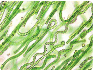
2. Photoheterotrophs: These organisms make use of light as energy source and organic compounds as electron and carbon source. Example: Purple and Green Non sulphur bacteria
3. Chemoautotrophs: These are ecologically important microorganisms. They oxidize inorganic compounds like nitrate, iron and sulphur to obtain energy and electrons.
4. Chemoheterotrophs: These organisms use organic compounds to satisfy their needs of energy, electron and carbon. (Table 6.2)
ased on carbon, energy and electron sources
ole carbon source
substances from other organisms
ergy
al energy source (Organic or Inorganic)
inorganic substances
compounds
| Carbon, Energ y and Electron sources | |
|---|---|
| Carbon sources | |
| Autotrophs | CO as sole carbon source2 |
| Heterotrophs | Organic substances from other organisms |
| Energ y sources | |
| Phototroph | Light energ y |
| Chemotrophs | Chemical energ y source (Organic or Inorganic) |
| Electron sources | |
| Lithotrophs | Reduced inorganic substances |
| Organotrophs | Organic compounds |
Table 6.2: Nutritional classes of Microorgan
Nutritional class Energy/Electron/Carb
Litho photoautotrophs
Light energy Inorganic e- donor CO2
Organo photoheterotrophs
Light energy Organic e- donor Organic carbon source
Ltiho chemoautotrophs
Inorganic chemical com energy source Inorganic e- donor CO2
Organo chemoheterotrophs
Organic compounds as electron and carbon sou
Photosynthesis
Photosynthesis is a process of capturing light energy and converting into chemical energy. The chemical energy produced in the form of ATP and NADPH is used to synthesise organic compounds (carbohydrates); to be used as food. This ability makes photosynthesis, a significant process taking place on earth.
a. Blood lake in Texas-the bl of purple sulphur bacteria. b. Giant tube worms seen i nutrients given by chemolith
a) b)
isms
on source Organisms
Cyanobacteria, Purple and Green sulphur Bacteria
Purple and Green Nonsulfur bacteria
pounds as Nitrifying bacteria, Iron bacteria
energy, rce.
Most pathogenic bacteria, fungi and protozoa.
Eukaryotes (plants and algae) and prokaryotes (cyanobacteria and purple, green bacteria) are capable of carrying out photosynthesis. Cyanobacteria perform photosynthesis in a similar manner to plants.
Photosynthesis in Bacteria There are four groups of photosynthetic bacteria. They are green sulphur bacteria
ood red colour is due to the excess presence
n deep sea hydrothermal vents survive on otrophic bacteria.
| Nutritional class | Energ y/Electron/Carbon source | Organisms |
|---|---|---|
| Lithophotoautotrophs | Light energ y Inorganic e donorCO-2 | Cyanobacteria, Purple and Green sulphur Bacteria |
| Organo photoheterotrophs | Light energ yOrganic e donorOrganic carbon source- | Purple and Green Nonsulfur bacteria |
| Ltihochemoautotrophs | Inorganic chemical compounds as energ y sourceInorganic e donorCO-2 | Nitrifying bacteria, Iron bacteria |
| Organo chemoheterotrophs | Organic compounds as energ y, electron and carbon source. | Most pathogenic bacteria, fungi and protozoa. |
(Example: Chlorobium) and green non sulphur bacteria (Example: Chloroflexus) purple sulphur bacteria (Example: Chromatium) and purple non sulphur bacteria (Example: Rhodospirillum). These photosynthetic bacteria can fix atmospheric CO2 in a similar fashion like cyanobacteria but using only one photosystem and using H2S as the electron donor instead of H2O.
Process of photosynthesis in bacteria The electron transport system in purple and green bacteria consists of only one Photosystem PSI (P870). They do not possess photosystem II. When P870 gets excited upon capture of light energy, it donates the electron to bacteriopheophytin. Electrons flow through quinones and cytochromes and are reverted back to P870. This process is cyclic (since the electron excited from P870 comes back to P870) and generates ATP. A reversed electron flow operates in purple bacteria to reduce NAD+ to NADH. Electrons are extracted from external electron donors like hydrogen sulphide, hydrogen, elemental sulphur and organic compounds to synthesise NADH. Since H2O is not used as electron donor, oxygen is not evolved which explains the anoxygenic nature of the organisms involved (Figure 6.3). The sulphur evolved during this reaction is deposited as sulphur globules either outside or inside the cells. CO2 + H2S (CH2O)n + S. Table 6.3 compares the photosystheic process in plants, algae and bacteria.
1. What will be the electron flow sequence of noncyclic and cyclic photo phosphorylation?
2. Chemical energy produced in photosynthesis is either ATP NADPH or ATP NADH. Why?
HOTS
Microbial Growth
In bacteria, growth can be defined as an increase in cellular constituents. Growth results in increase of cell number.
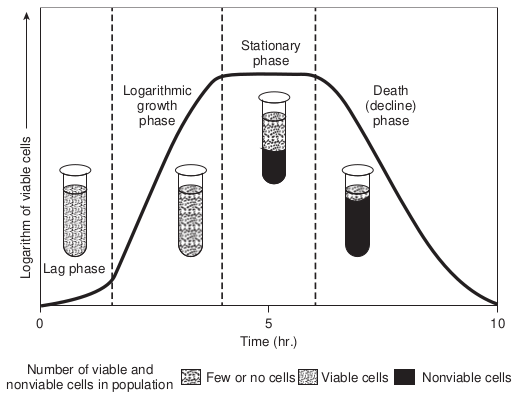
When bacteria are cultivated in liquid medium and are grown as batch culture (Growth occurring in a single batch of medium with no fresh medium provided), cell multiplication happens till all the nutrients are exhausted. After sometime, nutrient concentrations decline and bacterial cells begin to die. This growth pattern can be plotted in a graph as the logarithm of viable cells versus incubation time (Figure 6.4). The growth curve has four distinct phases.
1. Lag phase
2. Logrithmic phase/Exponential phase
3. Stationary phase
4. Death phase
1. Lag Phase When bacteria are introduced into fresh medium, no immediate cell multiplication and increase in cell numbers occur. The cell prepares itself for cell division by synthesizing cell components and increase in cell mass. Since there is a lag in cell division, this phase is called lag phase.
2. Logrithmic Phase/Exponential Phase During this phase, microorganisms rapidly divide and grow at a maximal rate possible utilizing all the nutrients present in the medium. The growth rate is constant during the exponential phase. The organism divides and doubles in number at regular intervals. The growth curve rises smoothly.
3. Stationary Phase As the nutrients get depleted, the cell growth stops and the growth curve become horizontal. The total number of viable cells remains constant which is due to a balance between cell division and cell death.
4. Death Phase Nutrient deprivation and build up of wastes lead to the decline in cell numbers. The microbial population dies rapidly and logarithmically and the growth curve also stops down.
Batch culture It is the growth of microorganisms in a fixed volume of culture medium in which nutrient supply is not renewed and wastes are not removed. It is a closed system. This can be used to study the various growth phases of microorganisms.
Continuous culture A continuous culture is an open system with constant volume to which fresh
Stat ph
Logarithmic growth phase
Lag phase
Number of viable and nonviable cells in population Few or no
0 Time
Lo ga
rit hm
o f v
ia bl
e ce
lls
medium is added and utilized (spent) medium are removed continuously at a constant rate. A microbial culture remains in exponential state for longer periods, for days and even weeks. This enables the researcher to learn about the physiological processes and enzymatic activities of organisms.
There are two ways by which continuous culture is operated.
a) Chemostat b) Turbidostat
Chemostat The chemostat operates so that the sterile nutrient medium enters the culture vessel at the same rate as the spent medium is removed. The chemostat can control growth rate and cell density simultaneously and independently of each other. Two factors play an important role in achieving this dilution rate and concentration of the limiting nutrient (a carbon or a nitrogen source like sugars or
Death (decline) phase
ionary ase
cells Viable cells Nonviable cells
5 (hr.)
10
phases of growth in laboratory conditions
|——|
|——|
|——|
|——|
| Lag phase |Logarithmicgrowthphase |Stationaryphase |Death(decline)phase |
aminoacids). Growth rate can be controlled b is controlled by modifying the concentration
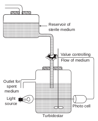
Turbidostat This type of continuous culture system has a photocell that measures the turbidity of the culture vessel. This automatically regulates the flow rate of the culture medium. Turbidostat does not contain limiting nutrients (Figure 6.6).
Factors Influencing Growth
The growth and activities of microorganisms are greatly influenced by the physical and chemical conditions of their environment. Among all factors, four key factors play major roles in controlling the growth of microorganisms. They are 1. Temperature 2. pH 3. Water activity 4. Oxygen
Fresh medium
Control valve
Air supply
Air filter
Culture vessel
Receptacle
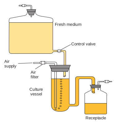
y adjusting the dilution rate and cell density of the limiting nutrient (Figure 6.5).
1. Temperature Temperature is one of the most important environmental factor affecting the growth and survival of microorganisms. Temperature can affect microorganisms because the enzyme catalysed reactions are sensitive to fluctuations in temperature.
For every microorganism, there is a minimum temperature below which no growth occurs, an optimum temperature at which growth is most rapid, and a maximum temperature above which no growth occur. These three temperatures are called cardinal temperatures.
Temperature classes of microorganisms Microorganisms are broadly distinguished into four groups in relation to their temperature optima.
• Psychrophiles
Light source
Turbidostar
Photo cell
Flow of medium Value controlling
Reservoir of sterile medium
Outlet for spent
medium
|——|——|——|
|——|——|——|
• Mesophiles • Thermophiles • Hyperthermophiles
Psychrophiles A psychrophile can be defined as an organism with an optimal growth temperature of 15°C, maximum growth temperature of 20°C and a minimum growth temperature at 0°C. These organisms are found in polar regions like Arctic and Antarctic oceans. They are rapidly killed as the temperature rises because the cellular constituents start to leak due to cell membrane disruption. Some examples of psychrophiles are Moritella, Photobacterium and Pseudomonas.
Snow alga – Chlam- ydomonas nivalis grows within the snow and its brilliant red coloured spores are
responsible for the formation of pink snow.
Why do unopened pasteurized milk spoil even under refrigeration?
HOTS
Psychrotolerant Organisms that can grow at 0°C, but have temperature optimum growth temperature range of 20°C-40°C are called psychrotolerant.
Mesophiles These are microorganisms that grow in optimum temperature between 20-45°C, they have a temperature minimum of 15-20°C and a maximum temperature of 45°C. All human pathogens are mesophiles.
Thermophiles Organisms whose growth temperature optimum is between 55-65°C are called thermophiles. They have minimum growth temperature of 45°C. These organisms are found in compost stacks, hot water lines and hot springs. They contain enzymes that are heat stable and protein synthesis systems function well at high temperature.
Taq polymerase, a DNA polymerase enzyme which is of great applied importance used in DNA amplification. It is isolated from Thermus aquaticus, a thermophile.
Infobits
Hyperthermophiles Organisms whose growth optimum temperature is above 80°C are called hyperthermophiles. These are mostly bacteria and archaebacteria. They are found in boiling hot springs and hydrothermal vents on seafloor.
2. pH pH is defined as the negative logarithm of the hydrogen ion concentration. pH scale extends from pH 0.0 to pH 14.0 and each exchange of 1 pH unit represents a 10 fold change in hydrogen ion concentration. pH greatly influences microbial growth. Each organism has a definite pH range and well defined pH growth optimum. Most natural environments have pH values between 5 and 9.
Organisms are classified into Acidophiles, Neutrophiles and Alkalophiles based on their optimum growth pH.
Acidophiles are organisms that grow best at low pH (0.0–5.5) Example: Most fungi, bacteria like Acidithiobacillus, Archaebacteria like Sulfolobus and Thermoplasma.
Neutrophiles are organisms that grow well at an optimum pH between 5.5 and 8.0. Most bacteria and protozoa are neutrophiles.
Organisms that prefer to grow at pH between 8.5-11.5 are called alkalophiles. These microorganisms are typically found in soda lakes and high carbonate soils. Example: Bacillus firmus.
3. Water Activity and Osmosis Water activity, (aw) is the ratio of vapour pressure of the solution to the vapour pressure of pure water (aw values vary between 0 and 1). Water activity is inversely related to osmotic pressure. Organisms that can grow in low aw values are called osmotolerant. Example: Staphylococus aureus.
Only a few organisms are capable of tolerating high salt concentration and still growing optimally in low water activity. Such organisms are called halophiles. Halophiles can grow in 1-15% Sodium chloride (NaCl) concentrations. Organisms that can grow in very salty environments are called extreme halophiles. (They can grow in 15-30%) NaCl concentration. Example: Halobacterium.
Crenation: Shriveling of cytoplasm in the cell is called crenation. This effect
helps to preserve some foods.
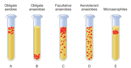
4. Oxygen Most of the microorganisms require oxygen for their optimal growth but some of them survive very well in total absence of oxygen and are killed when exposed to air.
Based on their need and tolerance for oxygen, microorganisms are classified into the following types.
(1) Obligate aerobes exhibit growth only at full oxygen level (21% O2 on air) because O2
is needed for their respiration and metabolic activities Example: Micrococcus, most Algae, Fungi and Protozoa.
(2) Microaerophiles are aerobes that require oxygen at levels lower than that of air. Example: Azospirillum, Campylobacter, Treponema
(3) Obligate anaerobes does not require oxygen for their respiration and growth. This group cannot tolerate O2 and are killed in its presence. Example: Methanogens, Clostridium.
(4) Aerotolerant anaerobes can grow in the presence of oxygen though O2 is not required for their growth. Example: Streptococcus pyogenes.
(5). Facultative anaerobes can grow either under oxic or anoxic conditions: Example: Escherichia coli. (Figure 6.7)
Measurement of Growth
Different methods are employed for measuring the cell growth of microorganisms. Cell growth is indicated by increase in the number of cells or increase in weight of cell mass. There are direct and indirect methods of measuring microbial growth.
1. Direct Measurements Total count and viable count are the two methods widely employed to count cell numbers.
Total count: The total number of cells in a population can be measured by counting a sample under the microscope. This is called direct microscopic count. This is done by using a specialized counting chamber called Petroff Hausser chamber which is a specially designed slide with a grid. The liquid sample is placed on the grid which has a total area of 1mm2 and divided into 25 large squares. The number of cells in large square is counted and the total number of cells is calculated by multiplying it with a conversion factor based on the volume of the chamber (Figure 6.8).
Advantages This is a quick method of estimating cell numbers.
Obligate aerobes
A B C
Obligate anaerobes
Facultat anaerob
0.05 mm
Count cells in this square
0.25 mm
(a) (b)

Disadvantages 1. Dead cells are also counted 2. Special microscopes like phase contrast
microscope are needed if unstained samples are used.
3. Small cells are difficult to count 2. Viable Count A viable cell is one that is able to divide and form a visible colony on the nutrient media. Viable cells are counted by methods pour plate and spread plate.
Pour plate method In this method, a known volume (0.1 or 1.0ml) of the culture is pipetted into a sterile petri plate, then molten nutrient medium is poured over and incubated. Colonies will appear throughout the agar medium and are counted to obtain viable count.
D E
ive es
Aerotolerant anaerobes Microaerophiles
e growth of various types of bacteria
1.00 mm
1.00 mm
1.00 mm
sser counting chamber tion of bacterial cells
|——|——|——|——|——|——|
|——|——|——|——|——|——|——|——|——|
Spread plate method In this method, a known volume of the culture (0 agar medium, using a sterile spreader. The total n incubation represents the total number of viable c 3. Measurement of Cell Mass A cell suspension appears turbid or cloudy due to this cell suspension, microbial cells scatter light s turbidity increases, more light is scattered and less amount of unscattered light can be measured usin indirectly related to cell numbers.
.1ml) is plated and spread over solidified sterile umber of colonies appearing on the plate after
ells in the culture.
active cell growth. When light is passed through triking them. As the concentration of cells and light is transmitted through the suspension. The
g a spectrophotometer, the values of which are
ICT CORNER
Step3
Step1
Cultur
URL: http://learn.chm.msu.edu/vibl/content/ differential.html
Preparation of Bacteriological Media
STEPS: • Use the URL or scan the QR code to re • Click module at the bottom and read • Follow the steps and open activities u
one and explore it. • Record your observation of Differen
specimen suitable for particular med
Step4
Step2
e Media
ach ‘Virtual Interactive Bacterial Laboratory’. the description and steps. nder’ Common Bacteriologic Media’ one by
tial Media. Click examples and record the ia
Summary Microorganisms need macro and micronutrients for their growth. Based on the energy source, organisms are grouped into Phototrophs and Chemotrophs. Based on carbon source, they are classified into autotrophs and heterotrophs. Organisms are grouped into lithotrophs and organotrophs based on their electron source. The four nutritional classes of microbes are photoautotrophs, Photoheterotrophs, chemoautotrophs and chemoheterotrophs.
Cyanobacteria are prokaryotes that can perform photosynthesis. Chlorophyll is the pigment needed to capture light energy (photons). In cyanobacteria and green plants, non cyclic photophosphorylation takes place to generate ATP and NADPH during photosynthesis whereas cyclic photophosphorylation takes place in purple and green bacteria involving only one Photosystem (PS I).
In a batch culture, bacteria show a characteristic growth pattern which consists of lag phase, log phase and stationary phase and decline phase. In a chemostat, cultures can be maintained in an exponential phase for long periods. The most important factors affecting microbial growth are temperature, pH and oxygen level. Total count and viable count are the two widely used methods to measure cell numbers.
Evaluation Multiple choice questions
1. An example of photoautotroph a. Cyanobacteria b. Algae c. Green plants d. All of the above
2. Magnesium is needed a. For cell wall synthesis b. As cofactor for enzymes c. For photosynthesis d. For protein synthesis
3. One of the following is an example for chemoautotroph a. Cyanobacteria b. Purple and green non sulphur
bacteria c. Iron bacteria d. Protozoa
4. The phase of growth in which the growth rate is equal to the death rate is a. Stationary phase b. Death phase c. Exponential phase d. Lag phase
5. Organisms that are capable of growing in 0°C are called a. Thermophiles b. Hyper thermophiles c. Barophiles d. Psychrophiles
6. Halophiles are organisms that can grow in a. Low water activity b. High salt concentration c. Low temperature d. High pH
7. An example of microaerophilic organism is a. Bacillus b. Azospirillum c. Pseudomonas d. Escherichia.coli
8. The specialized chamber used for the counting of microbial cells is a. Haemocytometer b. Counting chamber c. Petroff Hauss chamber d. Counting slide
Answer the following
1. Give notes on the nutritional classes of microorganisms.
2. Classify microorganisms based on energy and carbon source.
3. What are light and dark reactions in photosynthesis?
4. What is bacteriochlorophyll? Give its role.
5. Define chemoautotroph. 6. Define photosynthesis. 7. Give examples of photosynthetic
bacteria. 8. What do mean by cardinal temperature? 9. Give notes on photosynthetic pigments.
10. What are halophiles? 11. Give reason for the ability of
thermophiles to grow in high temperatures.
12. How bacterial cells are counted using counting chamber?
13. Classify microorganisms based on their temperature requirement.
Student Activity • Expose a container with water to sunl
cyanobacteria on water which explains th • Store a loaf of bread for a week after the
of fungi/molds which demonstrates the • Collect rusted iron pipes which contain ch
oxidize iron for their nutrients. • Place two bowls of cooked rice/vegetabl
another at room temperature at 30-35°C. rice stored at 30-35°C. Check the pH of m
14. Describe the role of macro and micronutrients in microorganisms. How do you think bacteria acquire their nutrients from their environment?
15. Explain the classification of microbes based on their nutrition. If H2S is toxic to living organisms, how do purple and green bacteria survive and use H2S in such environments.
16. Describe the photosystems of cyanobacteria.
17. Draw a schematic representation of Z scheme of non cyclic photophosporylation.
18. Compare photosynthesis between plants, cyanobacteria and purple green bacteria.
19. Explain the principle and uses of chemostat and turbidostat with diagrams.
20. Describe the classification of microorganism based on their oxygen requirement.
21. Explain the principle and uses of chemostat and turbidostat with diagrams.
22. Explain the relation of osmosis to water activity.
23. Define growth. Explain the phases of growth of bacteria with neat diagram.
ight for a week. Observe the growth of e photoautotrophic mode of nutrition. expiry date. You can observe the growth mode of nutrition of chemoheterotrophs. emolithotrophic Thiobacillus sp which can
es–one inside the refrigerator at 6°C, and Give reasons for the quick spoilage of the ilk using a pH paper.