After studying this chapter the student will be able,
• To describe the importance of medical microbiology.
• To understand the types and sources of infections.
• To know the types of infectious diseases and virulence factors of the pathogen.
• To tell the etiological agents of skin wound respiratory, gastro intestinal, ocular, urinary, reproductive, ner- vous system and systemic infections.
• To know the causative agents of varoius human diseases and their portal of entry.
Chapter 12 Medical Microbiology Chapter Outline
12.1 Microbial Infections of the Human Body
12.2 Skin and Wound Infections 12.3 Respiratory Tract Infections 12.4 Gastrointestinal Tract Infections 12.5 Ocular Infections 12.6 Urinary Tract Infections 12.7 Reproductive Tract Infections 12.8 Infections of the
Nervous System 12.9 Systemic
Infections
an tes inn inf
Learning Objectives
Medical Microbiology or Clinical Microbiology plays important role by providing the necessary diagnostic ting, means of epidemiological detection, and future ovation required in an era of emerging and reemerging
ectious diseases.
Microbial Infections of the Human Body
Medical microbiology is the branch of microbiology which deals with prevention, diagnosis and treatment of infectious diseases. There are four kinds of microorganisms that cause infectious diseases. They are bacteria, fungi, parasites and viruses. Any disease that spreads from one host to another, either directly or indirectly is said to be a communicable disease. Chicken pox, measles, genital herpes, typhoid fever and tuberculosis are examples of such diseases, that are easily spread from one person to another.
A non communicable disease does not spread from one host to another. For example, Clostridium tetani, a soil
|——| | Me dic a l M icr obio log y o r C linic a l M icr obio log y p l aysan im p or t ant r ole b y p rov idin g t he n e ces s ar y di ag nos t ictes t in g , m e ans o f ep idemio log ic a l det e c t io n, a nd f utur einn ova t io n re quir e d in an era of em er g in g and re em er g in ginf e c t io us di s e as es. |
inhabitant, produces Tetanus when it is introduced into a wound or an abrasion. Tetanus is thus an infectious disease, but not communicable.
Infectious disease occurs when the infecting microorganism causes damage to the host. The term infection refers to the establishment of the microorganisms in the tissues resulting in injury or harmful effect to the host. Infection is a pathological condition due to the growth of microorganisms in a host. To initiate an infection, a pathogenic microbe enters the tissues of the body by a characterization route, the portal of entry.
Routes of Infections
There are various ways in which microorganisms enters into the host are explained below.
a. Contact Infection may be acquired by contact which may be direct or indirect. Sexually transmitted diseases such as syphilis and gonorrhea spread by direct contact. Indirect contact may be through the agency of inanimate objects such as clothing, pencils or toys which may be contaminated by a pathogen from one person to another. Pencils shared by school children may act as fomites in the transmission of diphtheria, and face towels in trachoma.
b. Inhalation Respiratory infections such as influenza and tuberculosis are transmitted by inhalation of the pathogen in droplet and droplet nuclei that are shed by the patients during sneezing, speaking or coughing. Common cold virus, Adenovirus is
some of the virus producing respiratory infections.
c. Ingestion Intestinal infections are generally acquired by the ingestion of food or drinks contaminated by pathogens. Infection transmitted by ingestion may be waterborne (cholera), food borne (typhoid) or fecal-oral route (dysentery).
d. Inoculation Pathogens, in some instances, may be inoculated directly into the tissues of the host. Tetanus spores implanted in the depth of wounds, rabies virus deposited subcutaneously by dog bites, inoculation through unsterile syringes and surgical equipments are examples that enter through direct inoculation.
e. Congenital Some pathogens are able to cross the placental barrier and infect the fetus in uterus. Bacteria like Treponema pallidum, viruses like Rubella, Cytomegalovirus parasite like Toxoplasma gondi are some of the organisms that enter through placenta and cause disease in the newborn.
Types of Infections
Infections may be classified in various ways. Initial infection with a parasite in a host is called a primary infection. Subsequent infections by the same parasite in the host are termed reinfections. When a new parasite sets up an infection in a host whose resistance is lowered by a preexisting infectious disease, this is termed secondary infection.
When in a patient already suffering from a disease, a new infection is setup
from another host or other external sources it is termed cross infection. Cross infections occurring in hospitals are called nosocomial infections. Iatrogenic infection refers to physician induced infections resulting from investigative, therapeutic or other procedures.
Depending on whether the source of infection is from the host’s own body or from external sources, infections are classified as endogenous or exogenous, respectively.
Endogenous infection Endogenous infections are acquired from the host himself from the normal flora of the body.
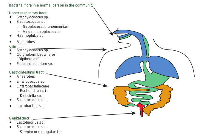
Microorganisms are present in certain areas of the body in all human beings. They are called normal flora. The common areas are Nose, Mouth, Teeth, Throat, Intestine, Urethra, Vagina and Skin (Figure 12.1).
Staphylococcus sp.
Bacterial flora in a normal person in the community
Upper respiratory tract
Skin
Gastrointestinal tract
Genital tract
Streptococcus sp. Streptococcus pneumoniae–
– Viridans streptococcus Haemophilus sp. Anaerobes
Staphylococcus sp. Coryneform bacteria or ‘‘Diptheroids’’ Propionibacterium sp.
Anaerobes
Streptococcus sp.
Enterococcus sp. Enterobacteriaceae – Escherichia coli
– Streptococcus agalactiae
– Klebsiella sp.
Lactobacillus sp.
Lactobacillus sp. Streptococcus sp.
1. When the skin is breached normal flora enters the tissues.
2. When the urethral organisms ascend, they cause urinary tract infection
3. When a patient is treated with antibiotics, normal flora is eliminated and replaced by potential pathogens
4. When the intestine is perforated, normal flora enter the previously sterile body parts
5. Similarly when the pH of the vagina increases potential pathogens occupy the space.
However normal flora helps host against pathogen and benefits the host in many ways • Normal flora of skin produces fatty
acids which inhibit other species • Intestinal bacteria secrete antibacterial
substances (bacteriocins, colicins) and many metabolic products that prevent other species to survive.
s present as normal flora n a site. Only few are listed)
• Because of their large numbers other species do not have space in the intestine
• Acidic environment created by vaginal Lactobacilli suppresses growth of other bacteria.
How do normal flora help host against pathogenic microorganisms?
HOTS
Exogenous sources of infections Human beings: The commonest source of infections in human are from other human beings. The parasite may originate from a patient or a carrier. A patient is a person who harbours the pathogenic microorganism and suffers from ill effect because of it. A healthy carrier is the one who harbours the pathogens but has never suffered from the disease caused by the pathogen. A convalescent carrier is one who has recovered from the disease and continues to harbor the pathogen in his body (Figure 12.2).
Animals: Many pathogens are able to infect both human beings and animals. Infectious disease transmitted from animals to human beings are called zoonoses. Zoonotic diseases may be bacterial (Example: plague from rats) or viral (Example: rabies from dogs).
Insects: Blood sucking insects may transmit pathogens to human beings. The diseases so caused are called arthopod borne diseases. Insects such as mosquitoes, ticks, mites, flies, fleas and lice that transmit infections are called vectors.
Transmission may be mechanical or biological. Mechanical transmission is the passive transport of the pathogens on the insects feet or other body parts. Example: Houseflies can transfer the pathogens of Typhoid fever and Bacillary dysentery
The story of typhoid Mary The classic example of role of carriers in disease transmission is the story of Mary Mallon.
Mary Mallon was an Irish immigrant who worked as a cook in New York in the early twentieth century. Over seven years, from 1900 to 1907, Mallon worked for number of different households. Unknowingly spreading illness to the people who lived in each one. Later George Soper, tracked Mallon linked 22 cases of typhoid fever through her. He discovered that Mallon was a carrier for typhoid but was immune to it herself. Although active carriers had been recognized before, this was the first time that an asymptomatic carrier of infected had been identified. Epidemiologists were able to trace 51 cases of typhoid fever and three deaths directly to Mallon, who is remembered as “Typhoid Mary”. She was forced to prison and then released under the conditions that she could no longer be a cook. She assumed a false name and began cooking again and of course, infecting numerous people. She was again prisoned where she died 26 years later of pneumonia.
Infobits
(shigellosis) from feces of infected people to food. Such vectors are called mechanical vectors.
Biological transmission is an active process and is more complex. The pathogens multiply in the body of the vectors often undergoing part of a developmental cycle in it. Such vectors are termed biological vectors. Example: Aedes aegypti mosquito transmitting dengue, Anopheles mosquito transmitting malaria.
Soil: Some pathogens are able to survive in the soil for very long periods. Spores of tetatus bacilli may remain viable in the soil for several decades and serve as the source of infection.
Water: Water may act as the source of infection due to contamination with pathogenic microorganisms. Example: Cholera causing Vibrio cholerae.
Food: Contaminated food materials may act as source of infection. The presence of pathogens in food may be due to external contamination. Example: Food contaminated by Staphylococcus.
Types of Infectious Diseases
Infectious diseases may be localised or generalised.

Localised infections: An infection that is restricted to a specific location or region within the body of the host is called localised infection.
Generalised infections: An infection that has spread to several regions or areas in the body of the host. This involves the spread of the infecting agent from the site of entry through tissue spaces or channels, along the lymphatics or through the bloodstream.
Circulation of bacteria in the blood is known as Bacteremia. Septicemia is the condition where bacteria circulate and multiply in the blood, form toxic products and cause high fever. Pyemia is a condition where pyogenic bacteria produce septicemia with multiple abscesses in the internal organs such as the spleen, liver and kidney.
Occurrence of a disease To understand the full scope of a disease, we should know about its occurrence. Epidemiology involves in the study of the frequency and distribution of disease and other health related factors in defined populations. The incidence of a disease is the number of people in a population who develop a disease during a particular time period. The prevalence of a disease
n a microbe causes disease in a host
is the total number of existing cases with respect to the entire population.
Depending on the spread of infectious disease in the community, they may be classified into different types.
• Endemic diseases are those which are constantly present in a particular area. Typhoid fever is endemic in most parts of India.
• Epidemic disease is one that spreads rapidly, involving many persons in an area at the same time. Example: Epidemic of Dengue in 2017.
• A pandemic is an epidemic that spreads through many areas of the world involving very large numbers of persons within a short period. Example: H1N1 Influenza outbreak in 2009. Ebola outbreak in 2014-2016 in West Africa was the largest in history and first ever epidemic, affecting multiple countries.
• If a particular disease occurs only occasionally, it is called a sporadic disease. The most commonly occurring sporadic diseases in India are Diphtheria and Hepatitis A and E.
Severity or duration of a disease Another useful way of defining the scope of a disease is in terms of its severity or duration. • An acute disease is one that develops
rapidly but lasts for a short time. • A chronic disease develops more
slowly, and the body’s reactions may be less severe, but the disease is likely to be continual or recurrent for long periods.
• A disease that is intermediate between acute and chronic is described as a subacute disease.
Facts about Fever: Fever is as more healthful than harmful. An experiment with vertebrates shows that fever increases the rate of antibody synthesis. Increased temperatures
stimulate the activities of T cells and increase the effectiveness of interferon. Fever appears to enhance
phagocytosis. Fever almost never occurs as a single response; it is usually accompanied by chills. The explanation lies in the natural physiological interaction between the thermostat in the hypothalamus and the temperature of the blood. For example: If the thermostat has been set (by pyrogen) at 102°F but the blood temperature is 99°F, the muscles are stimulated to contract involuntary (shivering) as a means of producing heat. In addition, the vessels in the skin constrict, creating a sensation of cold and the piloerector muscles in the skin develops ‘goose bumps’.
• A latent disease is one in which the causative agent remains inactive for a time but then becomes active to produce symptoms of the disease.
Interaction between Microbes and Host
Pathogen is a microorganism which causes disease.
Pathogenecity is the ability of a pathogen to produce disease.
Virulence is the degree of pathogenecity of a microorganism. Virulence is not generally attributable to a single property
but depends on several parameters related to the organism, the host and their interaction.
Microorganisms first enter the body, survive, multiply and elaborate many factors and produce the disease.
Adhesion: The initial event in the pathogenesis of many infections is the attachment of the bacteria to body surfaces. Adhesions may occur as organized structures, such as fimbriae and pili. Adhesions serve as virulence factors.
Capsule: It is an envelope or slime layer surrounding the cell wall of certain microorganisms. Capsule plays important roles in immune evasion as it inhibit’s phagocytosis, as well as protecting the bacteria while outside the host.
Toxins: Toxins are specific chemical products of microbes, plants and some animals that are poisonous to other organisms. Toxigenicty is the power to produce toxins.
A toxin is named according to specific target of action: Neurotoxin acts on the nervous system. Enterotoxin acts on the
Table 12.1: Differences between endotoxin a
Exotoxins Heat labile proteins, secreted by certain species of bacteria and diffuse readily into the surrounding medium Proteins with a strong specificity to a target cell and extremely powerful sometimes deadly Highly immunogenic Toxoids can be made by treating toxins with formalin Produced mainly by Gram positive bacteria but also by some Gram negative bacteria
intestine, Haemotoxin lyses red blood cells, and Nephrotoxins damages the kidneys.
A toxin molecule secreted by a living bacterial cell into the infected tissue is an exotoxin. A toxin that is not actively secreted but is shed from the outer membrane is an endotoxin. the difference between exotoxin and endotoxin were given in Table 12.1.
Production of enzymes Some enzymes like proteases, DNAases and phospholipases are produced and they help in destruction of the cell structure and to hydrolyse host tissues.
Antigenic variation Microorganisms evade the host immune responses by changing their surface antigens. Antigenic drift and antigenic shift are common in influenza viruses. The distinction between the commensal and the organism associated with disease.
Diagnostic Cycle
Specific diagnosis is important for better patient care, use of appropriate antibiotics
nd exotoxin
Endotoxins Heat stable polysaccharide proteins, lipid complex which form an integral part of the cellwall of Gram negative bacteria A Lipopolysaccharide (LPS), which is part of the outermembrane of gram negative cell walls Less immunogenic Toxoids cannot be made
Produced by Gram negative bacteria
| E xotoxins | E nd otoxins |
|---|---|
| He at l abi le p rotein s, s e cr et e d b y cer t ainsp e cies o f b ac ter i a a nd dif f us e r e adi ly in tot he s ur roun din g m e di um | He at s t able p olys acc har ide p rotein s, li pidco mplex w hic h f or m a n in teg ra l p ar t o f t hece l lwa l l o f G ra m n ega t ive b ac ter i a |
| Protein s w it h a s t rong s p e cif ici t y t o a t argetce l l and ext rem ely p ower f u l s omet im esde ad ly | A L ip op olys acc har ide (LPS), w hic h i s p ar tof t he o uter mem bra ne o f g ra m nega t ivece l l wa l ls |
| Hig h ly imm un og enic | L es s imm un og enic |
| Toxoid s c an b e m ade b y t re at in g t oxin sw it h f or ma lin | Toxoid s c ann ot b e m ade |
| Pro duce d main ly by Gra m p osi t ive bac ter i abut a ls o b y s ome G ra m n ega t ive b ac ter i a | Pro duce d b y G ra m n ega t ive b ac ter i a |
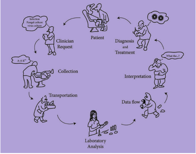
and to initiate appropriate preventive measures. The diagnostic cycle begins when the clinician takes a microbiological sample and ends when a clinician receives the laboratory report and uses the information to manage the condition (Figure 12.3).
The steps in diagnostic cycle are
1. Clinical request and provision of clinical information.
2. Collection and transport of appropriate specimens.
3. Laboratory analysis. 4. Interpretation of microbiology report
and use of the information. Specimen Collection and transport:
It is important to collect the specimen appropriately and protect it from contamination. Transport media are used that are compatible with the organism
believed to be present in the clinical sample. Quality of patient specimens and their transport to the laboratory is important.
Infections and samples used Respiratory tract infections: Nasal and bronchial washings, throat and nasal swabs, sputum.
Eye infections: Conjunctival swab or scraping.
Wound infections: Pus, skin scraping, wound swap.
Gastrointestinal infections: Stool, rec- tal swabs.
Genital infections: Vesicle fluid or swab.
Urinary tract infections: Urine. Blood borne infections: Blood. Nervous system infections: Cerebro-
spinalfluid (CSF).
s in diagnostic cycle
Laboratory diagnosis of infectious agents Direct diagnosis: It is the
demonstration of the presence of an infectious agent, antigen or nucleic acids
Indirect diagnosis: It is the demonstration of presence of antibodies to a particular infectious agent, cytopathic effects, haemagglutination, inclusion bodies and neutralization.
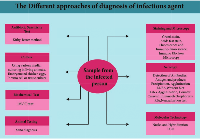
The different approaches for diagnosis or identification of infectious agents are shown in Figure 12.4.
Skin and Wound Infections
The skin, which covers and protects the body, is the body’s first line of defense against pathogens. As a physical barrier, it is almost impossible for the pathogens to penetrate it. However, microorganisms
can enter through skin breaks that are not readily apparent, and the larval forms of a few parasites can penetrate the intact skin. The skin has up to seven layers (Figure 12.5) of ectodermal tissue and guards the underlying tissues viz; muscles, bones, ligaments and internal organs. Nearly all human skin is covered with hair follicles. Because it interfaces with the environment, skin plays an important role in protecting the body against pathogens and excessive water loss. Its other functions are insulation, temperature regulation, sensation, synthesis of vitamin D, and the protection of vitamin B folates. Severely damaged skin will try to heal by forming scar tissue. This is often discolored and depigmented.
pproaches of diagnosis
Normal Microbiota of the Skin
The skin’s normal microbiota contains relatively large numbers of Gram positive bacteria, such as Staphylococci and Micrococci. Bacteria in the skin tends to be grouped into small clumps. Vigorous washing can reduce their numbers but will not eliminate them. Microorganisms remaining in hair follicles and sweat glands after washing will soon reestablish the normal populations. Areas of the body with high moisture, such as armpits and between the legs, have higher populations of microorganisms. They metabolize secretions from the sweat glands and are the main contributors to body odour.
Also part of the skin’s normal microbiota are Gram positive pleomorphic rods called diphtheroids. Some diphtheroids, such as Propionibacterium acnes, are typically anaerobic and inhabit hair follicles. These bacteria produce propionic acid, which helps maintain the low pH of skin, generally between 3 and 5
Wound Infection
Wound can be defined as any interruption of continuity of external or internal surfaces caused by violence
Wounds may occur following: surgery, trauma or injections
Wound infections may occur mainly after surgical procedures
Wound sepsis is the result of cross infection from human sources and from other outside sources.
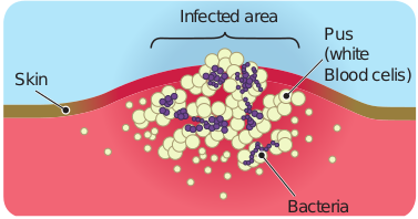
Bacteria associated with wound infections Many bacteria are associated with wound infection (Figure 12.6). The normal flora
may also cause infection. The most common normal flora of the skin are: Staphylococci, and various Streptococci, Sarcina sp, anaerobic Diphtheroids, Gram negative rods and others.
Pus (white Blood celis)
Infected area
Bacteria
Skin
Post operative infections Gasgangrene organisms like Clostridium perfringens, Staphylococcus aureus and Clostridium tetani may cause post operative infections.
1. What are the possible infecting agent you could pick up when you are injured while playing on the ground? List them and name the diseases that they could cause.
2. What are the possible infectious agent that can infect you when you are injured by a rusted nail?
HOTS
Route of entry Wounds may occur following surgery, trauma or injections. Wound infections may occur mainly after surgical procedures. Wound sepsis is the result of cross infection from human sources and from other outside sources. Infections of skin are listed in Table 12.2.
Mechanisms of damage 1. Organisms enter through the skin,
multiply there and produce the disease in the skin. For example, impetigo, abscess and cellulitis (Figure 12.7) are caused by Staphylococcus aureus and Streptococcus pyogenes.
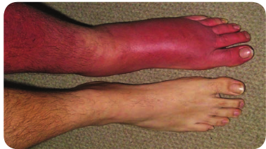
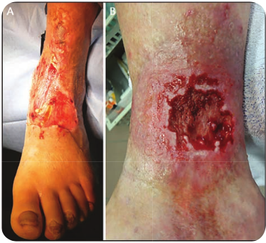
As soon as the organisms enter the skin they multiply and produce various toxins that kill the cells and produce cellulitis. Further damage leads to necrosis and ulcer formation (Figure 12.8).
Table 12.2: Bacterial Infections of the skin
Disease Pathogen Si
Cellulitis Streptococcus pyogenes
Loca of d hypo warm the t
Erysipelas Streptococcus pyogenes
Infla of sk may
Impetigo Staphylococcus aureus, Streptococcus pyogenes
Vesi som nose
Wound infections Pseudomonas aeruginosa, others
Form or o
2. Organisms multiply in the skin and produce disease in internal organs. For example some Group A Streptococci multiply in the skin and produce disease known as Acute Glomerulonephritis causing damage to the kidneys. Some times Corynebacterium diphtheriae may multiply in the skin and affect the heart due to the toxin
gns and Symptoms Transmission
lised inflammation ermis and
dermis; skin red, , and painful to
ouch
Through cut or abrasion
med, swollen patch in, often on face;
be suppurative
Through cut or abrasion
cles, pustules, and etimes bullae around and mouth
Highly contagious, especially via contact
ation of biofilm in n wound
Exposure of wound to microbes in environment; poor wound hygiene
| Dis e as e | Patho ge n | Sig ns a nd S y mptoms | Tr ans miss i on |
|---|---|---|---|
| C ell ul iti s | Strep toc oc c u spyogen es | L o c a li s e d inf l amm at io nof der mi s a ndhyp o der mi s; s k in r e d,wa r m, a nd p ainf u l t ot he t ouc h | Thr oug h c ut o r a bra sio n |
| Er ysi p el as | Strep toc oc c u spyogen es | Inf l ame d, sw ol len p atchof s k in, o f ten o n face;may b e s uppura t ive | Thr oug h c ut o r a bra sio n |
| Imp et ig o | Staphyloc oc c u saureu s, S treptococcu spyogen es | Vesic les, p ustu les, a nds omet im es b u l l ae a roun dno s e a nd mo ut h | Hig h ly co nt ag io us,es p e ci a l ly v i a co nt ac t |
| Woun d inf e c t io ns | Ps e udom on a saer ug inos a, ot he rs | For mat io n o f b io f i lm inor o n w oun d | E xp os ur e o f w oun dto micr ob es inenv ir onm en t; p o orwoun d h yg ien e |
3. Sometimes organism may multiply in the skin and produce the toxin which affect the Central Nervous System (CNS) and the effects seen. In the case of Clostridium tetani infection, convulsions and paralysis occur due to the production of a powerful toxin.
Respiratory Tract Infections
With every breath, we inhale several microorganisms and therefore the respiratory system is a major portal of entry for pathogens. In fact, respiratory system infections are the most common type of infections and among the most damaging. Some pathogens that enter via respiratory route can infect other parts of the body, such as skin incase of measles, mumps and rubella.
The upper respiratory system has several anatomical defenses against airborne pathogens. Coarse hairs in the nose, filter large dust particles from the air. The nose is lined with a mucous membrane that contains numerous mucous secreting cells and cilia. The upper portion of the throat also contains a ciliated mucous membrane. The mucous moistens inhaled air and traps dust and microorganisms. The cilia help to remove these particles by moving them towards the mouth for elimination.
Structure of Respiratory Tract
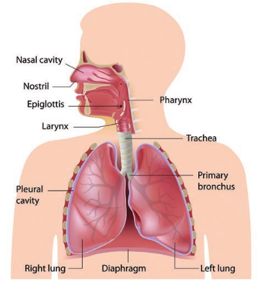
The structure of respiratory tract is divided into two main parts viz: upper respiratory tract (URT) and lower respiratory tract (LRT).
Upper respiratory tract includes mouth, nose, nasal cavity, sinuses, throat or pharynx, epiglottis and larynx.
Lower respiratory tract includes trachea, bronchi, bronchioles, lungs and alveoli (Figure 12.9).
Normal Defenses against Infections
Respiratory tract infection are divided into upper respiratory tract (URT) tract infection and lower respiratory tract (LRT) infection. Infection of the respiratory tract are listed in the Table 12.3.
URT: Infections are Sinusitis, Pharyn- gitis Laryngitis and Epiglotitis
LRT: Infections are Trachiitis, Tracheo bronchitis, Bronchitis, Alveolitis and Pneumonia.
Upper respira Diseases Pathogen
Bacterial Epigottitis Haemophilus influenzae Streptococcal pharyngitis (strep throat)
Streptococci, especially Streptococcus pyogenes
Diphtheria Corynebacterium diphth
Otitis media Several agents, espe Staphylococcus au Streptococcus pneumoni Haemophilus influenza
Viral di Common cold Rhino virus
Lower respira Bacterial
Pertussis (whooping cough)
Bordetella pertussis
Tuberculosis Mycobacterium tubercu
Viral di Respiratory syncytial virus (RSV)
Respiratory syncytial vi
Fungal d Blastomycosis Blastomyces dermatitidi
Bacterial pn Pneumococcal pneumonia
Streptococcus pneumoni
Haemophilus influenzae pneumonia
Haemophilus influenzae
tory system Symptoms
diseases Inflammation of the epiglottis Inflamed mucous membranes of the throat;
eriae Bacterial exotoxin interferes with protein synthesis; damages heart, kidney, and other organs; membrane forms in throat; cutaneous form also occurs;
cially reus,
a and
Accumulations of pus in middle ear build up painful pressure on eardrum
seases Familiar symptoms of coughing, sneezing, running nose.
tory system diseases
Cilia in upper respiratory tract inactivated, mucus accumulates, spasms of intense coughing to clear mucus;
losis Tubercle bacilli entering lungs survive phagocytosis, reproduce in macrophages; tubercles formed to isolate pathogen; defenses eventually fail, and infection becomes systemic;
seases rus A serious respiratory disease of infants;
iseases s Abscesses; extensive tissue damage; eumonia
a Infected alveoli of lung fill with fluids; interferes with oxygen uptake Symptoms resemble pneumococcal pneumonia
| Upp er r es pir ator y s ystem | ||
|---|---|---|
| Dis e as es | Patho ge n | Sy mpt om s |
| Bac teri a l dis e as es | ||
| Epi gott it is | Haem ophilu s i nf luenz ae | Inf l amm at io n o f t he ep ig lo tt is |
| St rep to co cc a lphar y ng it is (s t rept hr o at) | Strep toc oc c i , es p e ci a l lyStreptococcu s p yogen es | Inf l ame d muco us mem bra nes of t het hr o at; |
| Di pht her i a | C or y nebac te r ium d iphthe r iae | B ac ter i a l ex otoxin in ter fer es w it hprotein sy nt hesi s; d amages h e ar t,k idn e y, a nd o t her o rga ns; m em bra nefor ms in t hr o at; c ut ane ous f or m a ls oo cc ur s; |
| Oti ti s m ed i a | S e vera l a gen ts, es p e ci a l lyStaphylococcu s a ureu s,Streptoco cc u s pne umo nia and Haem ophilu s i nf luenz a | Acc um u l at io ns o f pus in midd le e arbui ld u p p ainf u l p res sur e o n e ardr um |
| Vir a l dis e as es | ||
| C omm on co ld | R hin o v ir us | Fami li ar sy mptoms o f co ug hin g ,sn e e zin g , r unnin g n os e. |
| L owe r r e sp i r ator y s yst em | ||
| Bac teri a l dis e as es | ||
| Per tussi s (w ho opin gco ug h) | B ordetella p e r tu ssi s | Ci li a in upp er res pira tor y t rac tin ac t iva te d, muc us acc um u l ates, sp asm sof in ten s e co ug hin g t o c le ar m uc us; |
| Tub er c u losi s | Myc oba c te r ium t ube rc ulo si s | Tub er cle b aci l li en ter in g l un gssur v ive p hago c yt osi s, r ep ro duce inmacr ophages; t ub er cles f or me d t ois ol ate p at hog en; def en s es e ven tu a l lyfa i l, a nd inf e c t io n b e co mes sys temic; |
| Vir a l dis e as es | ||
| R es pira tor y sy nc yt i a lv ir us (RSV ) | R es pira tor y sy nc yt i a l v ir us | A s er io us r es pira tor y di s e as e o f infa nts; |
| Fun g a l dis e as es | ||
| Bl astomycosi s | Bla stomyc es de r matitid i s | Abs ces s es; ext en si ve t issue d amage; |
| Bac teri a l p ne umo ni a | ||
| Pneum o co cc a lpneum oni a | Streptoco cc u s p ne umo nia | Inf e c te d a lve oli o f l un g f i l l w it h f luid s;in ter fer es w it h o xyg en u pt a ke |
| Haem ophi lusinf luenzae p neum oni a | Haem ophilu s i nf luenz ae | Sy mptoms r es em ble p neum o co cc a lpneum oni a |
Gastrointestinal Tract Infections
Human systems function by the energy produced from the digested food molecules. The food is swallowed through mouth and digested in the gastro intestinal tract. The food we consumed should be free of contaminations. The contaminated food causes gastrointestinal infections.
Through contaminated food and water the pathogens are ingested and they enter the GIT. In the small intestine they initiate an infection. Many times the pathogens that cause intestinal infections multiply in the GIT and produce their pathogenic effect in the intestine itself. Example: Shigellosis, Cholera.
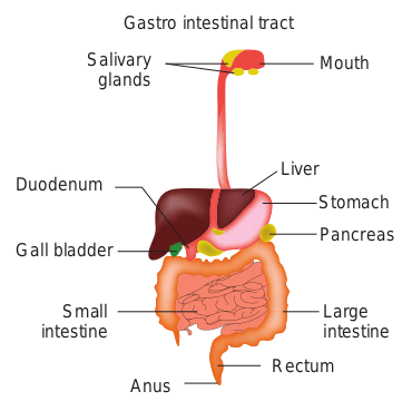
The gastrointestinal tract (GIT) or alimentary canal includes the mouth, pharynx, throat, oesophagus (food Tube lead to the stomach), stomach, small and large intestine. It also includes accessory structures salivary glands, liver, gall bladder and pancreas lying outside the GIT. Secretions of these organs enhance the digestion of food molecules (Figure 12.11).
MouthSalivary glands
Gastro intestinal tract
Stomach
Liver Duodenum
Gall bladder
Small intestine
Anus Rectum
Pancreas
Large intestine
Difference between infection and intoxication Microbial diseases of digestive system are typically transmitted by a fecal oral route. Most such diseases result from the ingestion of food or water contaminated with pathogenic microorganisms or their toxins. These pathogens usually enter the food or water supply after being shed in the feces of people or animals infected with them.
• Each milliliter of saliva can contain millions of bacteria
• Stomach/small intestine has very few microorganisms because of hydrochloric acid present in the stomach.
• Large intestine harbours microbial population exceeding 100 billion of bacteria per gram of feces (40% fecal masses contain microbial cell material)
• Large intestine microbial population mainly contain anaerobes and facultative anaerobes.
After ingestion of pathogenic microorganisms, localization and multiplication of organisms takes place in the GIT and is called infection. Microorganisms may penetrate into intestinal mucosa and grow there or they may penetrate to other organs. Gastroenteritis is usually classified as either infection or intoxication. Food borne diseases can arise from either infection or intoxication. In both cases, bacterial toxins are typically responsible for producing disease signs and symptoms. In a food infection the microbial agent ingested
colonise in the gut and then produces toxins that damage host cells.
In case of food intoxication the toxins produced by bacteria in the food are ingested. Infection and intoxication differ in their onset of symptoms. Infections are characterized by a delay in the appearance of gastrointestinal disturbance until the pathogen increases in number or affects invaded tissue. Infection is correlated with onset of fever, one of the basic body’s general responses to an infective organism. In case of intoxication, the symptoms are characterized by sudden appearance of gastrointestinal disturbances like cramping, nausea, vomiting or diarrhoea.
What is likely to happen to a child who drinks contaminated water?
HOTS
Microbial Flora of Gastrointestinal Tract
The stomach and gastrointestinal tract are not sterile and are colonized by the organisms that perform functions beneficial to the host, including the manufacture of essential vitamins. Escherichia coli found in the intestine help the body to produce vitamin K and Bifodobacteria can synthesizes vitamins such as vitamin B12, folate, and riboflavin. Humans cannot produce these vitamins. The normal flora changes according to the diet, age, cultural conditions and the use of antibiotics (Table 12.4).
Terms used in GIT Infections
Gastroenteritis: Inflammation of lining of stomach and intestine. It is a syndrome
characterized by nausea, vomiting, diarrhea, abdominal discomfort.
Stomach is acidic because of the pres- ence of hydrochlo- ric acid. So in this
acidic condition organisms gen- erally not survive except one bac- terium Helicobacter pylori. This bacterium is the leading cause of stomach ulcers. This bacterium has maximum evidence of correlation with the development of stomach and intestinal cancer.
Stomach
H. pylori
Diarrhea: Condition in which feces, are discharged from the bowels frequently and in a liquid form.
Botulism is a special case of intoxication because, the ingestion of the preformed toxin
affects the nervous system rather than GIT.
Infant Botulism is the infectious form of Botulism which results when spores of Clostridium botulinum swallowed colonise in the intestine. Botulism spores can be found in honey.
Dysentery: Inflammatory disorder of the G
Gastritis: Inflammation of the stomach lin
Enteritis: Inflammation of the intestinal m
Colitis: Inflammation of the colon
Hepatitis: Inflammation of the liver
Enterocolitis: Inflamation involving the mu
Peritonitis: Inflammation of peritoneum lining of the abdominal cavity). Infections of
Table 12.5: Diseases of the digestive system
Infection Pathogen
Bacterial Staphylococcal food poisoning
Staphylococcus aure
Shigellosis (bacillary dysentery)
Shigella sp-
Salmonellosis salmonella enterica Typhoid fever Salmonella typhi
Cholera Vibrio Cholerae Yersinia gastroenteritis Yersinia enterocoliti
Viral Di Mumps Mumps virus
Paramyxoviridae Viral gastroenteritis Rotavirus
Fungal D Ergot poisoning Claviceps purpurea
Aflatoxin poisoning Aspergillus flavus
Table 12.4: Normal flora of human gastroint
At human birth Stoma Breast fed babies Lactob Bottled milk fed babies Enter
Clostr Small intestine Lactob Large intestine Anaer
Bifido
IT associated with pus and blood in feces.
ing that results in swelling.
ucosa
cosa of both large and small intestine.
(it is the serous membrane that forms the digestive system are listed in Table 12.5.
Symptoms
Diseases us Nausea, vomiting, and diarrhea
Tissue damage and dysentery
Nausea and diarrhea High fever, significant mortality
Diarrhea with large water loss ca Abdominal pain and diarrhea,
usually mild; may be confused with appendicitis
seases Painful swelling of parotid glands
Vomiting, diarrhea for 1 week iseases
Restricted blood flow to limbs; hallucinogenic
Liver cirrhosis; liver cancer
estinal tract
ch and intestine are sterile acillus bifidus
ic bacteria, Lactobacillus bifidus, Enterococci, idium sp acilli, Enterococcus faecalis, Escherichia coli obic bacteria, Streptococci, Bacteroides, bacteriumbifidum
| At h um an b ir t h | Stomac h a nd in tes t in e a re s ter i le |
|---|---|
| Bre ast f e d b abies | L ac tob ac illu s b if idu s |
| B ott le d mi l k f e d b abies | En ter ic bac ter i a, L ac tob ac illu s bif id us, Enter ococci,C lostr idium sp |
| Sma l l in tes t in e | L ac tobaci lli, Enter ococcu s faecali s, E s cher ichia coli |
| L arge in tes t in e | Anaer obic b ac ter i a, Strep toc oc c i , B ac te roi des ,Bif idob ac te r iumbif idum |
| In fe ct io n | Patho ge n | Sy mpt om s |
|---|---|---|
| Bac teri a l Dis e as es | ||
| St aphy lo co cc a l f o o dp oi s on i ng | Staphyloc oc c u s a ure u s | Naus e a, v omi t in g , a nd di ar rhe a |
| Shig el losi s (b aci l l ar ydys en ter y) | Shige lla s p- | Tissue d amage a nd d ys en ter y |
| Sa lm onel losi s | s almo nella e nte r ica | Naus e a a nd d i ar rhe a |
| Typ hoid f e ver | S almon ella t y phi | Hig h f e ver, sig nif ic ant m or t a li t y |
| C holera | Vibr io Ch oler ae | Di ar rhe a w it h l arge wa ter los s |
| Yer sini a ga st ro en ter it is | Ye rs inia e nte roc olitic a | Ab do min a l p ain a nd di ar rhe a,usu a l ly mi ld; m ay b e co nf us e d w it happ en dici t is |
| Vir a l Dis e as es | ||
| Mum ps | Mumps v ir u sParamy x ov ir idae | Painf u l sw el lin g o f p arot id g l ands |
| Vira l ga st ro en ter it is | Rotav ir u s | Vomi t in g , di ar rhe a f or 1 w e ek |
| Fun g a l Dis e as es | ||
| Er got p ois onin g | C lav ic ep s p ur purea | R es t r ic te d b lo o d f lo w t o lim bs;ha l lucin og enic |
| Af l atoxin p ois onin g | Aspe rg ill u s f lav u s | L iver cir rhosi s; li ver c ancer |
Ocular Infections
A number of microorganisms cause infection when introduced into the mucosa of the eye. In general, bacterial eye infections can lead to inflammation, irritation, and discharge, but they vary in severity. Some are typically short-lived, and others are chronic and lead to permanent eye damage. Prevention requires limiting the exposure to contagious pathogens. When infections do occur, prompt treatment with antibiotics can often limit or prevent permanent damage.
The external surfaces of the eye viz. the conjunctiva and cornea are susceptible to infection. These are exposed to external world and are easily accessible to infective agents. Particularly the conjunctiva is susceptible because it is covered with eyelid that provides warm, moist and enclosed environment in which contaminating organisms can quickly establish a focus of infection. However, eyelid and tears
Table 12.6: Bacterial infections of the eye
Disease Pathogen S Acute bacterial conjunctivitis
Haemophilus influenza
In co p
Bacterial keratitis Staphylococcus epidermidis, Pseudomonas aeruginosa
R o se p sc le
Neonatal conjunctivitis
Chlamydia trachomatis, Neisseria gonorrhoeae
In co d an co b
igns and Symptoms Transmission flammation of njunctiva with
urulent discharge
Exposure to secretions from infected individuals
edness and irritation f eye, blurred vision, nsitivity to light;
rogressive corneal arring, which can ad to blindness
Exposure to pathogens on contaminated contact lenses
flammation of njunctiva, purulent
ischarge, scarring d perforation of rnea; may lead to
lindness
Neonate exposed to pathogens in birth canal of mother with chlamydia or gonorrhea
Infected eyelid and conjunctiva
Eyelid margin
Infected eyelid
Pus accumulation
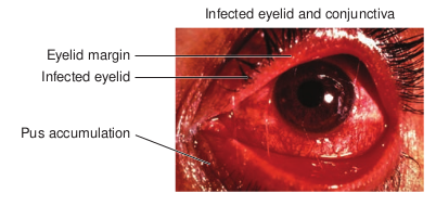
protect the external surfaces of the eye, both mechanically and biologically. Infection of Eyelid Most common cause of eyelid infection is Staphylococcus aureus.
Infection involves lid margins and cause blepharitis.
When the eyelid glands or follicles are affected stye (sticky eye) is seen (Figure 12.14).
Conjunctivitis (inflammation of conjunctiva) Conjunctivitis or pink eye can be caused by many different kinds of viruses and bacteria.
| Dis e as e | Patho ge n | Sig ns a nd S y mptoms | Tr ans miss i on |
|---|---|---|---|
| Ac ute b ac ter i a lc onju nc t iv it is | Hae mo philu sinf lu e n z a | Inf l amm at io n o fco njun c t iva w it hpur u len t di s charge | E xp os ur e t os e cr et io ns f rominf e c te d in di v id u a ls |
| B ac ter i a l k era t it is | Staphyloc oc c u sep ide r mid i s , Ps e udom on a sae r ug ino s a | R e dn es s and irr it at io nof e ye, b lur re d v isio n,s en si t iv it y t o lig ht;prog res si ve co r ne a ls c ar r in g , w hic h c anle ad t o b lin dn es s | E xp os ur e t op at hog en s o nco nt amin ate dco nt ac t len s es |
| Neo nat alc onju nc t iv it is | C hla myd iatrac ho mati s , Nei ss er iag ono r rho eae | Inf l amm at io n o fco njun c t iva, pur u len td is charge , s c ar r i ngand p er fora t io n o fco r ne a; m ay le ad t oblin dn es s | Ne onate exp os e d t op at hog en s in b ir t hc ana l o f m ot herw it h c h l amydi a o rgono r rhe a |
Trachoma
Trachoma, or granular conjunctivitis, is a common cause of preventable blindness that is rare in the United States but widespread in developing countries, especially in Africa and Asia. The condition is caused by the same species that causes neonatal inclusion conjunctivitis in infants, Chlamydia trachomatis. Chlamydia trachomatis can be transmitted easily through fomites such as contaminated towels, bed linens, and clothing and also by direct contact with infected individuals. Chlamydia trachomatis can also spread by flies that transfer infected mucous containing Chlamydia trachomatis from one human to another. Infections of eye are listed in Table 12.6. ## Urinary Tract Infections The urinary system is composed of organs that regulate the chemical composition and the volume of the blood excrete mostly nitrogenous wastes products and water.
Many of the bacteria which cause UTI’s have developed resistance to antibiotics. Research has turned to probiotic (Lactobacillus) strain which stimulates immune function, lowers acidity levels in the urinary tract, and discourages the growth of UTI causing organisms.
Infobits
Table 12.7: Microorganisms involved in UTI
Microorganisms Bacteria (most common) Escherichi coli, Viruses Adenovirus, M Fungi Trichomonas va Parasites Candida
The urinary system consists of two kidneys, two ureters, a single urinary bladder and a single urethra. Wastes are removed from the blood as it circulates through the kidneys (Figure 12.15).
Ureter
Bladder
Urethra
LO W
ER T
RA C
T U
PP ER
T RA
C T Kidney
Ureteritis (Ureter infection)
Pyelonephritis (Kidney infection)
Cystitis (Bladder infection)
Urethritis (Urethra infection)
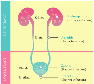
Infections of the kidney, ureter and bladder constitute Urinary Tract Infections (UTI). When infection occur in the kidney and ureter it is called upper urinary tract infections and bladder downwards is called lower urinary tract infections. Urinary tract infection is common in females than males. The urinary system normally contains few microbes but it is subjected to opportunistic infections that can be quite troublesome. Almost all such infections are caused by bacteria although occasional infections by pathogens such as parasites, protozoa and fungi also occured. Microorganisms invloved in UTI are listed in Table 12.7.
Examples Klebsiella, Enterobacter, Proteus umps ginalis, Schistosoma haematobium
| Mi cr o org anis ms | E xa mp l es |
|---|---|
| B ac ter i a (m os t co mm on) | E s cher ichi c oli, K lebsiella, E nter obac ter, P roteu s |
| Vir us es | Aden ov ir us, M um ps |
| Fu ng i | Tr icho mo na s v ag ina li s , S chi stos oma hae matobium |
| Para si tes | C andida |
Predisposing Factors for UTI
Urinary tract infection is common in females than in males. The urethra in females are shorter and wider and is less effective in preventing the bacteria entering the bladder. Sexual intercourse is a predisposing factor in females. High incidence is seen in pregnant women due to hormonal changes and impairment of urine flow due to pressure on urinary tract.
Obesity increases the risk of UTI’s in men. A 2013 study examineed
how obesity affected the chance of developing UTI and it was found that obese men were twice more likely to develop the UTI than obese women.
Urinary Tract Infection caused by Escherichia coli
Escherichia coli is the predominant cause of UTI.
It is a normal flora of the gut and can cause extra intestinal infections (UTI, Wound infection.) UTI (it can also be
Table 12.8: Microbial Diseases of the Urinar
Disease Patho Bacterial Diseases of the Urinary system Cystitis (Urinary bladder infection)
Escherichia col Staphylococcus saprophyticus
Pyelonephritis (Kidney infection)
Primarily Esch
Leptospirosis (Kidney infection)
Leptospira inte
involved in other infections like wound infection peritonitis) UTI is common in (a) married women (b) elderly men with prostate enlargement.
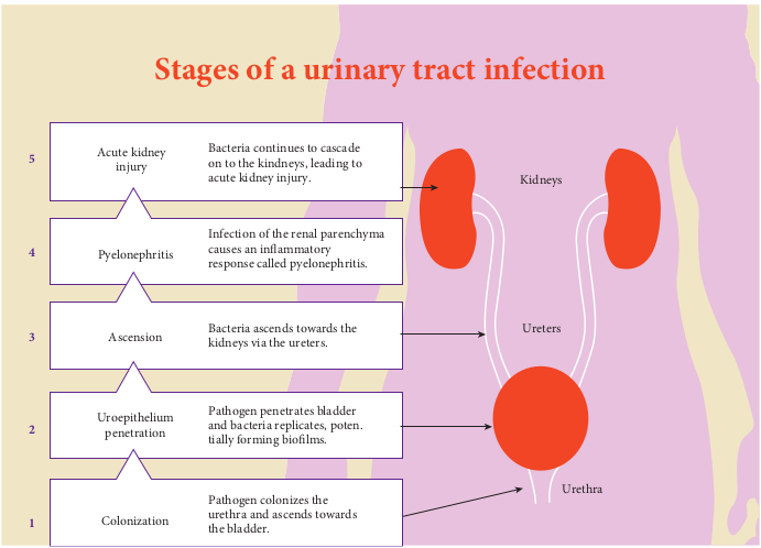
Pathogenesis of cystitis in woman Bladder infections can result from the downward migration of organisms from an infected kidney. But majority arise by ascent of pathogens from the rectum and vagina to the urethra meatus and bladder, leading to cystitis. If left untreated, the infection can further ascend to involve the kidneys (pyelonephritis) (Figure 12.16).
The rectum and vagina function as the reservoir of bacteria for sporadic infections
In men, the longer urethra is believed to protect against ascending infections.
When Escherichia.coli (and other Gram Negative rods) causes UTI, usually the number of organisms in freshly passed urine is more than 100,000 organisms/ml.
This is called “significant bacteriuria”. Counts less than this is associated with contaminants from urethra or externalia. Infection of urinary tract are listed in Table 12.8.
y System
gen Symptoms i_,_
Difficulty or pain in urination
erichia coli Fever; back or flank pain
rrogans Headaches, muscular aches, fever; kidney failure a possible complication
| Dis e as e | Patho ge n | Sy mpt om s |
|---|---|---|
| B ac ter i a l Di s e as es o f t heUr in ar y sys temCys t it is (U r in ar y b l adderinf e c t io n) | E s ch e r ichia co li , Staphyloc oc c u ss aprophy tic u s | Dif f ic u lt y o r p ain inur in at io n |
| P yelo nep hr it is (K idn e yinf e c t io n) | Pr i mar i ly E s ch e r ichia c oli | Fe ver ; b ac k o r f l an k p ain |
| L ep tos pir osi s (K idn e yinf e c t io n) | L eptospira i nter roga ns | He ad ac hes, m us c u l ar ac hes,fe ver ; k idn e y fa i lur e ap os si ble co mplic at io n |
Reproductive Tract Infections
Reproductive tract infections are caused by organisms normally present in the reproductive or genital tract or introduced from the outside during sexual contact or medical procedures. It occur both in men and women. Based on mode of infection reproductive tract infections are classified into three types:
1. Sexually Transmitted Disease
It is caused through means of sexual contact. Examples: Chlamydia, Gonnorhea, Chancroid, and Acquired Immuno Deficiency Syndrome (AIDS).
2. Endogenous Infections
These are caused by the overgrowth of
Infection of the renal parenchyma causes an in ammatory response called pyelonephritis.
Bacteria continues to cascade on to the kindneys, leading to acute kidney injury.
Acute kidney injury5
4
3
2
1
Pyelonephritis
Stages of a urinar
Ascension
Uroepithelium penetration
Colonization
Bacteria ascends towards the kidneys via the ureters.
Pathogen penetrates bladder and bacteria replicates, poten. tially forming bio lms.
Pathogen colonizes the urethra and ascends towards the bladder.
organisms normally present in the genital tract of healthy women. Example: Bacterial Vaginosis or Vulvo Vaginal Candidiasis.
3. Iatrogenic Infections
These infections are associated with improperly performed medical procedures such as unsafe abortion or poor delivery practices. The endogenous organisms in the vagina or sexually transmitted organisms in the cervix may be transferred during a transcervical procedure into the upper reproductive tract and cause serious infections of the uterus, fallopian tubes, and other pelvic organs.
In men reproductive tract infections transmitted by sexual contact are much more common than by endogenous or iatrogenic reproductive infections. In women reproductive infections spread through non sexual routes are usually more common.
Kidneys
Ureters
Urethra
Bladder
y tract infection
of a urinary tract infection
Mode of Transmission
Reproductive tract infections are caused by pathogenic bacteria, parasite, virus. It is mainly caused by pathogens entering into the body through the mucous mem- branes during unprotected vaginal, oral, anal intercourse with an infected part- ner. In developing countries bacterial infections like Gonorrhoea, Chlamydia, Syphilis, Bacterial Vaginosis, Lympho- granuloma Venereum, Trichomoniasis, Chancroid, and viral infections caused by Human Papilloma Virus, Hepatitis B Virus, Herpes Simplex Virus, Human Im- munodeficiency Virus are very common.
Normal Flora of Reproductive Tract
Mycobacterium smegmatis, a harmless commensal found in the smegma of the genitalia of both men and women. In nomal men aerobic and anaerobic bacteria, lactobacilli, alpha haemolytic Streptococci, Chalmydia trachomatis and Ureaplasma urealyticum may also be present.
The adult female genital tract has a very complex microflora. The character of the population changes with the variation of the menstrual cycle. Mostly the predominant bacteria are acid tolerant Lactobacilli. Glycogen is accumulated in the vaginal wall due to ovarian hormonal activity. The breakdown of glycogen by the lactic acid bacteria (Doderlien’s bacillus) leads to the formation of acidic pH (4.4-4.5). This acidic nature prevents the vagina from bacterial vaginosis and yeast infections. However before puberty and after menopause there is no glycogen formation. The normal
flora during this period contain normal skin microorganisms. The vaginal pH is mild alkaline. The normal vaginal flora often includes Listeria, anaerobic Streptococci, Mycoplasma, Gardnerella vaginalis, Neisseria, Spirochetes, Candida, Staphylococcus epidermidis.
Pathogenesis
After the entry of pathogenic organisms, with sufficient incubation time, symptoms are clearly manifested in the affected individual. The most common symptoms include unusual vaginal discharge, penile discharge, pelvic pain, itching, abnormal or heavy vaginal bleeding, rashes, warts, lesions, burning or pain during urination. However most of the infections are asymptomatic, which act as a effective control of reproductive tract infections. Diseases of reproductive system are listed in Table 12.9.
Tamilnadu has AIDS testing centres at all district head quarters with more than 55 Anti Retroviral Therapy(ART) centres and 750 (ICTC)-Integrated (voluntary) and confidential counselling and testing centres under the national AIDS control programme at district level government hospitals and medical colleges across the state.
Infobits
Table 12.9: Microbial diseases of the reprodu
Disease Pathogen
Bacterial
Gonorrhea Neisseria gonorrhoeae
P m f
Nongonococcal urethritis (NGU)
Chlamydia trachomatis or other bacteria, including Mycoplasma hominis and Urea plasma urealyticum
P C
Syphilis Treponema pallidum I r s a
Lymphogranuloma venereum (LGV)
Chlamydia trachomatis
S
Viral Di
Genital Herpes Herpes simplex virus type 2; HSV type 1
P
Genital warts Human papilloma viruses
W
AIDS Human Immunodeficiency virus (HIV)
l p ( f s
Fungal D
Candidiasis Candida albicans S d
Protozoan
Trichomoniasis Trichomonas vaginalis
V
ctive system
Symptoms
Diseases
ainful urination, discharge of pus in ales, abnormal vaginal discharge in
emales
ainful urination and watery discharge, hronic abdominal pain in females
nitial sore at site of infection, later skin ashes and mild fever; final stages may be evere lesions, damage to cardiovascular nd nervous systems.
welling in lymph nodes in groin
seases
ainful vesicles in genital area
arts in genital area
oss of appetite, weight loss, ersistent cough, attack on T cells immunocompromise), easily prone to ungal and other bacterial pathogens as econdary opportunistic infections.
iseases
evere vaginal itching, yeasty odor, yellow ischarge
Diseases
aginal itching, greenish yellow discharge
| Dis e as e | Patho ge n | Sy mpt om s |
|---|---|---|
| Bac teri a l Dis e as es | ||
| G ono r rhe a | Nei ss er iag ono r rho eae | Painf u l ur in at io n, di s charge o f p us inma les, a bnor ma l va g in a l di s charge infem a les |
| Non gon o c o c c a lur et hr it is (N GU) | C hla myd iatrac ho mati s o r o t herb ac ter i a, in cludin gMycopl a sm a h omini san d Urea pl a sm aurealy tic um | Painf u l ur in at io n a nd wa ter y di s charge,C hr onic a b do min a l p ain in f em a les |
| Syp hi li s | Trepo ne ma pa llid um | Ini t i a l s ore a t si te o f inf e c t io n, l ater s k inra shes a nd mi ld f e ver ; f in a l s t ages m ay b es e ver e lesio ns, d amage t o c ardio va s c u l arand n er vous sys tem s. |
| Ly mphog ra nu lo maven er eum (L GV ) | C hla myd iatrac ho mati s | Swel lin g in l y mph n o des in g roin |
| Vir a l Dis e as es | ||
| G eni t a l H er p es | Her pes s implex v i r us t yp e 2; HSV t yp e 1 | Painf u l v esic les in g eni t a l a re a |
| G eni t a l wa r ts | Hum an p api l lo mav ir us es | War ts in g eni t a l a re a |
| AIDS | HumanImm un o def icien c yv ir us (HIV ) | los s o f a pp et ite, w eig ht los s,p er si sten t co ug h, a tt ac k o n T ce l ls(imm un o co mpromi s e), e asi ly p rone t of un ga l a nd o t her b ac ter i a l p at hog en s a ss e co nd ar y o pp or tuni st ic inf e c t io ns. |
| Fun g a l Dis e as es | ||
| C andidi asi s | C andida a lbic an s | S e ver e va g in a l i tchin g , y e ast y o do r, y el lo wd is charge |
| Protoz o an Dis e as es | ||
| Tr ic homoni asi s | Tr icho mo na svag in ali s | Vag in a l i tchin g , g re eni sh y el lo w di s charge |
Infections of the Nervous System
Some of the most devastating infectious diseases are those that affect the nervous system, especially the brain and the spinal cord. Damage to these areas can lead to deafness, blindness, learning disabilities, paralysis and death. Microbial infections of CNS are infrequent but often have serious consequences. In pre antibiotic times, they were almost always fatal. An infection of CNS can be life threatening condition, especially for children with weakened immune system. These infections need quick diagnosis and immediate treatment by an infectious disease specialist. Bacteria, Fungi and viruses are the most common causes of CNS infections.
Structure of Nervous System
The human nervous system is organized into two divisions: The Central Nervous System (CNS) and Peripheral Nervous System (PNS). The Central Nervous System (CNS) consists of brain and
Venous blood
Ventricles
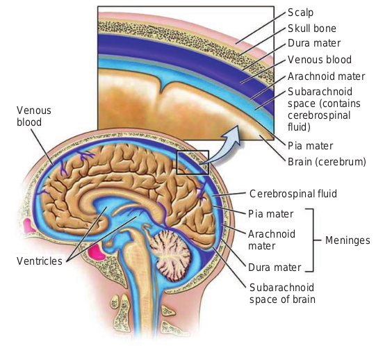
spinal cord. It controls most functions of the body and mind. The peripheral nervous system (PNS) consists of all the nerves that branch off from the brain and spinal cord. These peripheral nerves are the lines of communication between the CNS, the various parts of the body and the external environment (Figure 12.19).
Brain and spinal cord are covered by three layers of membranes called meninges. These layers are the outermost dura mater, the middle arachnoid mater, and the innermost pia mater. Between the pia mater and arachnoid membranes is a space called the subarachnoid space, in which there is cerebrospinal fluid (CSF) circulating.
Barriers of CNS
Dyes such as Trypan blue injected into the systemic circulation stain virtually all tissues, with the exception of the brain and spinal cord. This blood brain
f central nervous system
Scalp Skull bone Dura mater Venous blood Arachnoid mater Subarachnoid space (contains cerebrospinal fluid)
Pia mater Brain (cerebrum)
Cerebrospinal fluid
Pia mater
Arachnoid mater
Dura mater
Meninges
Subarachnoid space of brain
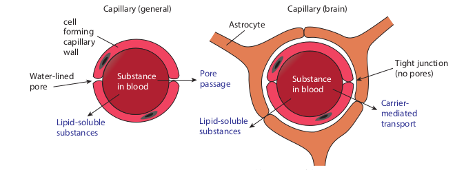
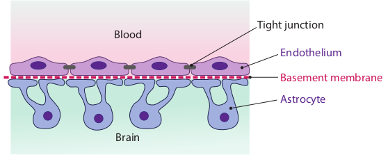
barrier excludes most macromolecules, microorganisms, immunocompetent cells and antibodies. Even pathogens that are circulating in the bloodstream usually cannot enter the brain and spinal cord because of blood brain barrier. Certain capillaries permit some substances to pass from the blood into the brain but restricts others. These capillaries are less permeable than others within the body and are therefore more selective in passing materials (Figure 12.20). The blood brain barrier (Figure 12.21) is due to the cellular
cell forming capillary wall
Substance in blood
Astr
Capillary (general)
Water-lined pore
Lipid-soluble substances
Pore passage
Lipid-solubl substances
Blood
Cerebrospinal uid
Blood
Brain
pillaries of brain
Substance in blood
ocyte
Capillary (brain)
e
Carrier- mediated transport
Tight junction (no pores)
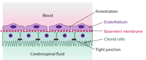
configuration of cerebral capillaries, the choroid plexus and arachnoid cells. It acts as a natural barrier that prevents the invasion of microorganisms into the brain. If this is breached organisms enter the brain. The blood CSF barrier (Figure 12.22) (also calle brain CSF barrier) consists of endothelium with fenestrations, and tightly joined choroid plexus epithelial cells. It acts as a natural barrier that prevents the invasion of microorganisms into the meninges.
od CSF barrier
Basement membrane
Endothelium
Fenestration
Tight junction
Chorid cells
od brain barrier
Basement membrane
Endothelium
Astrocyte
Tight junction
Routes through which microorganisms enter nervous system
• Skull or bone fractures
• Medical procedures
• Peripheral nerves
• Blood or lymph
Clinical Manifestations of Nervous System Infections
Some of the symptoms of nervous System infections are headache, fever, stiff neck, focal signs, seizures, confusion, weakness, hallucinations, stupor, coma, abnormal behavior and sleep disorder
Infections of Nervous System
• Meningitis is an inflammation of the meninges (membrane covering the brain). Meningitis is a diffuse infection caused by a variety of different agents.
• Encephalitis is defined as inflammation of the brain. Unlike an abscess, which is a localised area of bacterial or fungal growth, Encephalitis is usually due to
Drugs cannot cross the b soluble. Glucose and many can cross the barrier thro soluble antibiotic Chloramp
is only slightly lipid soluble, but, if it is tak the barrier to be effective. Inflammations barrier in such a way as to allow antibiotics there were no infection.
Antibodies found in the normal CNS present at low levels compared to serum le complement is also largely excluded. CSF many of the defenses found in the blood, s the microorganisms to enter CNS but it ham
viruses that produce more widespread intracellular infections.
• Brain abcess is a focus of purulent infection and is usually due to bacteria. Brain abscesses develop from either a contiguous focus of infection (such as the ears, the sinuses, or the teeth) or hematogenous spread from a distant focus (such as the lungs or heart, particularly with chronic purulent pulmonary disease, subacute bacterial endocarditis, or cyanotic congenital heart disease). In many cases the source is undetectable.
Etiological agents of Meningitis
This can be caused by a wide range of microorganisms and can be classified as pyogenic and non pyogenic meningitis. In pyogenic meningitis infiltration of pus cells (neutrophils) will be seen. In Non pyogenic or aseptic meningitis infiltration of lymphocytes may be seen. Diseases of nervous system are listed in Table 12.10.
lood brain barrier unless they are lipid amino acids are not lipid soluble, but they ugh special transport systems. The lipid henicol enters the brain readily. Penicillin en in very large doses, enough may cross of the brain tend to alter the blood brain to cross that would not be able to cross if
are derived from the serum and are vels. There are a few phagocytic cells and is especially vulnerable because it lacks uch as phagocytic cells. It is not easy for pers their clearance once it is penetrated.
Systemic Infections
An infection that is in the bloodstream is called a systemic infection. Systemic diseases such as flu and typhoid affect the entire body. Bacteria can enter the circulatory and lymphatic systems through acute infections or breaches of the skin barrier or mucosa. Breaches may occur through fairly common occurrences, such as insect bites or small wounds. Even the act of tooth brushing, which can cause small ruptures in the gums may introduce bacteria in to the circulatory system. In most cases, the Bacteremia
Table 12.10: Microbial diseases of the Nervo
Diseases Pathogen P Bacterial
Haemophilus influenzae meningitis
Haemophilus. Influenzae
R t
Meningococcal meningitis
Neisseria meningitidis
R t
Pneumococcal meningitis
Streptococcus pneumoniae
R t
Tetanus Clostridium tetani S Botulism Clostridium
botulinum M
Viral Di Poliomyelitis Poliovirus M
Rabies Lyssavirus, includ- ing rabies virus
S
Fungal D Cryptococcosis Cryptococcus
neoformans R r
Protozoan African trypanosomiasis
Trypanosoma brucei Rhodesiense, Trypanosoma brucei gambiense
S
result from such common exposure is transient and remains below the threshold of detection. In severe cases, bacteremia can lead to septicemia with dangerous complication such as Toxemia sepsis and Septic shock. In these situations, it is often the immune response to the infection that result in the clinical signs and symptoms rather than microbes themselves.
Summary
In the branch of medical microbiology we discussed about prevention, diagnosis and
us system
ortal of Entry Method of Transmission Diseases espiratory
ract Endogenous infection; aerosols
espiratory ract
Aerosols
espiratory ract
Aerosols
kin Puncture wound outh Food borne intoxication
seases outh Ingesting contaminated
water (fecal oral route) kin Animal bite
iseases espiratory
oute Inhaling soil contaminate with spores
Diseases kin Tsetse fly
| Dis e as es | Patho ge n | Por ta l o f E ntr y | Me tho d o f Tr ans miss i on |
|---|---|---|---|
| Bac teri a l Dis e as es | |||
| Haem ophi lusinf luenzae m enin g it is | Hae mo philu s .Inf luenz ae | R es pira tor yt rac t | En dog en ous inf e c t io n;aer os ols |
| Menin go co cc a lmenin g it is | Nei ss er iame ning itidi s | R es pira tor yt rac t | Aer os ols |
| Pneum o co cc a lmenin g it is | Strep toc oc c u spne umo niae | R es pira tor yt rac t | Aer os ols |
| Tet anus | C lostr idium t etani | Sk i n | Pun c tur e w oun d |
| B otu li sm | C lostr idiumbotulinum | Mout h | Fo o d b or ne in toxic at io n |
| Vir a l Dis e as es | |||
| Polio myeli t is | Polio v ir us | Mout h | Inges t in g co nt amin ate dwa ter (f e c a l o ra l r oute) |
| R abies | Lyssavir us, in clud -ing ra bies v ir us | Sk i n | Anim a l b ite |
| Fun g a l Dis e as es | |||
| Cr yp to co ccosi s | Cr y ptoco cc u sneofor man s | R es pira tor yroute | In ha lin g s oi l co nt amin atew it h s p ore s |
| Protoz o an Dis e as es | |||
| Af r i c an t r yp anos omi asi s | Tr y pano s omabr uce iR hode sie n s e ,Tr y pano s omabr ucei ga mbiens e | Sk i n | Ts ets e f ly |
treatment of infectious diseases. Infections are acquired through contact, inhalation, ingestion, inoculation and congenital. Sources of infections are endogenous and exogenous in origin. Normal flora are organisms present in certain areas of the body. Infectious diseases may be generalised or localised. Based on the occurrence of infectious diseases the infection may be epidemic, endemic, or sporadic. There are various virulence factors which are responsibility for the pathogenicity.
Skin is the first line of defence against pathogen. Normal uninterrupted skin provides protection against “invasion by bacteria”. Many exogenous and endogenous factors are responsible for wound infections. The mechanism of damage may be in the skin or some cases its spreads to the internal organs and CNS system.
Respiratory system of both lower and upper is the major path for entry of pathogens. The infections of upper respiratory tract are sinusitis, pharyngitis, laryngitis and epiglottitis. The infection of lower respiratory tract are trachitis, tracheobronchitis, bronchitis, and alveolitis.
Gastrointestinal tract infections are infections of the digestive system. The food borne infection and food intoxication are the common cause of gastroenteritis. The gut flora and natural defence mechanism by defensins bacteriocins, globet cells, IgA antibodies protect the individual against pathogenic infection. Diarrhea, dysentery, vomitting are the common symptoms of GIT. Oral rehydration therapy, proper hygiene to be manifested to reduce the risk of gastroenteritis.
The external structure and parts of the eye are easily susceptible to infections. The eyelids, tears, lysozyme, IgA are the natural
defence against infections. Conjunctivitis and Trachoma are the common eye diseases. Proper diagnosis and treatment should be suggested.
Uninary tract infections are more common in females than in males. There are many predisposing factors making female prone to the infections. The predominant causative agents in urinary tract infection is Escherichia.coli. The number of organisms in freshly passed urine is more than 100,000 organisms/ml. It is called significant bacteriuria.
The infections spread through reproductive tract by direct contact is called sexually transmitted disease. Mostly these infections are asymptomatic in women.
Nervous system infection affect brain and spinal cord. They are of two types meningitis and encephalatis. An infection that is in the blood stream is called systemic infections. Systemic diseases like flu and typhoid affect the entire body.
Modes of
Infection
STEPS: • Use the link or scan the QR code given
You can select any topic you wish. For • “Understanding Colds” page will open
CAT scan view etc.. • At the top left of the page click on “Me • Also select “Special features” and go
how penicillin kills bacteria in the “C penicillin against bacteria.
ICT CORNER
Step2Step1
Respiratory T
URL: https://www.cellsalive.com/toc_micro.htm
Know the myths of cold
below. “Cells Alive” home page will open. example click “understanding colds” . You can go through anatomy of the nose,
nu” and select “Treatments” and analyze. through the topic. Also you can select ells Alive” page, and know the action of
Step4Step3
ract Infections
Evaluation
Multiple choice questions
1. Syphilis is disease a. Sexually
transmitted disease b. Respiratory tract disease c. Gastro tract disease d. Urinary tract disease
2. is the person who harbours the pathogenic microorganisms and suffers from till effect because of it? a. Carrier b. Healthy carrier c. Patient d. All the above
3. Circulation of bacteria in the blood is known as a. Septicimia b. Pyemia c. Bacterimia d. None of the above
4. From the skull down to the brain, select the arrangement of layers of meninges from the following: a. Dura mater/Arachnoid mater/Pia
mater * b. Arachnoid mater/Dura mater/Pia
mater c. Pia mater/Arachnoid mater/Dura
mater d. Dura mater/Pia mater/Arachnoid
mater 5. Cerebrospinal fluid (CSF) is present
in which of the following? a. Perivascular spaces b. Sub arachnoid space *
c. Between skull and dura mater d. Sub dural space
6. antibody gives first line defense against respiratory tract infections. a. IgM b. IgA c. IgD d. IgE
7. The nose is lined with membrane.
a. Mucous b. Epithelial c. Secretion d. None of these
8. nature of stomach act as a natural defense mechanism. a. Acidic b. Neutral c. Alkaline d. None of the above
9. Traveller”s diarrhea is caused by a. Escherichia coli b. Staphylococcus aureus c. Vibrio cholerae d. All the above
10. is the predominant cause of UTI? a. Staphylocous aureus b. Escherichia coli c. Salmonella d. Streptococcus pyogenes
11. fungi involved in urinary tract infection? a. Klebsiella b. Candida sp c. Penicillium d. Escherichia coli
12. During the breakdown of glycogen by la as . a. Acidic b. Neutral c. Alkaline d. None of the above
Answer the following
1. Define congenital infection?
2. What is meant by nosocomial infection?
3. Define the term bacteremia, septicemia p
4. Explain mode of transfer of infection?
5. Define a wound.
6. What are the causes of wound?
7. Name two types of CNS infections.
8. Give the names of the etiologic agents of
9. Describe microbial disease of upper resp
10. State the difference between dysentery an
11. What is the difference between food born
12. Give the normal flora of the gastrointesti
13. What is called significant bacteriuria?
14. Explain the predisposing factors for urin
15. Define iatrogenic infection.
16. Explain the role of lactobacilli in the prev
17. Give detailed study of various bacteria reproductive tract infection.
ctobacilli in the vagina, makes vaginal pH
yremia?
wound infection.
iratory tract infection?
d diarrhoea?
e infection and intoxication?
nal tract of humans?
ary tract infection?
ention of bacterial vaginosis.
l, fungal and viral infectious diseases of
Student Activity (1)
1. Get information from your parents/neig to contamination. Example: If you drink
No After certain activity Gettin
1 Contaminate drinking water
Diarrh
2 3 4 5
2. Give a list of organisms present as norma is given in the text book).
3. Prepare model of respiratory tract with in Prepare a list of URT infections with the Observe a chronic smoker. He coughs ver collect information from nearby neigh immunized DTP vaccinated?
No Kid’s name DOB I 1 2 3
4. Student is asked to prepare a model of G See for example: What all the organisms that can be transm Give a list.
5. (1) Write an assignment on Madras eye ( (2) Write Dos and Don’s when a dust par
6. 1) Draw the structure of urinary tract in a the parts (make a poster presentation to urethra).
7. Prepare a chart showing all sexually tran Collect the disease photographs from the
8. Write a chart showing differences betwee
hbor about types of diseases one gets due contaminated water, you get diarrhoea.
g a disease Preventive method to advocate
oea Don’t drink or Boil, cool and drink
l flora of the skin (include other than that
novations. etiologic agents and prevention. y often. List out the reasons for his cough. bors kids (10). How many of them are
mmunized on Where corporation
or pvt
IT with their innovations.
itted through the fly contaminated food.
conjunctivitis due to viruses) ticle comes into your eye. chart board using your innovation. Label material with flow of urine from kidney
smitted diseases. net. n pyogenic and aseptic meningitis.
| No | Aft er c er ta in ac tiv ity | G e ttin g a dis e as e | Pre ventiv e me tho d t oadv o c ate |
|---|---|---|---|
| 1 | C ont am i natedr in k in g wa ter | D i ar rho e a | D on’t dr in k o r B oi l, co oland dr in k |
| 2 | |||
| 3 | |||
| 4 | |||
| 5 |
| No | K i d’s na me | D OB | Imm uniz e d o n | Wh ere c or p or ati onor p v t |
|---|---|---|---|---|
| 1 | ||||
| 2 | ||||
| 3 |